

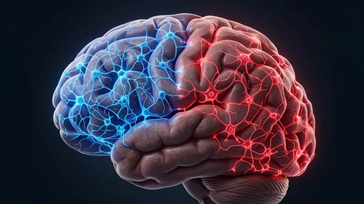
The research tracked the development of embryonic cells and found that regions linked to thought and vital functions take different paths from the earliest stages of the organ’s formation.
A study by Stanford Medicine has challenged a traditional concept regarding brain development. The work found that, from the earliest stages of embryonic development, two distinct groups of cells follow separate paths to form different regions of the organ: one gives rise to the forebrain—associated with functions such as language, thought, and decision-making—while the other forms the hindbrain, which is involved in vital processes like breathing, heart rate, sleep, and swallowing.
The study was published in the journal Nature Neuroscience and relied primarily on experiments with mouse embryos, alongside the analysis of human stem cells and comparisons with other species.
This finding does not mean that people literally have “two” independent brains. Rather, it suggests that the organ may have a more divided embryonic origin than previously thought. According to the researchers, distinct brain regions do not arise from a single type of precursor cell that later acquires different characteristics; instead, they stem from two cell populations that follow their own paths from a very early stage.
The pivotal moment is gastrulation, a phase of embryonic development during which cells begin to organize and receive instructions determining which tissues and organs they will form.
The team, led by Stanford researcher Kyle M. Loh, identified two cell populations. One is associated with the expression of the Otx2 gene and gives rise to the forebrain and midbrain. The other expresses Gbx2 and ultimately forms the hindbrain.
In the mouse embryos analyzed, the researchers observed that the two groups did not mix and maintained distinct trajectories throughout development.
Enviar comentarios

